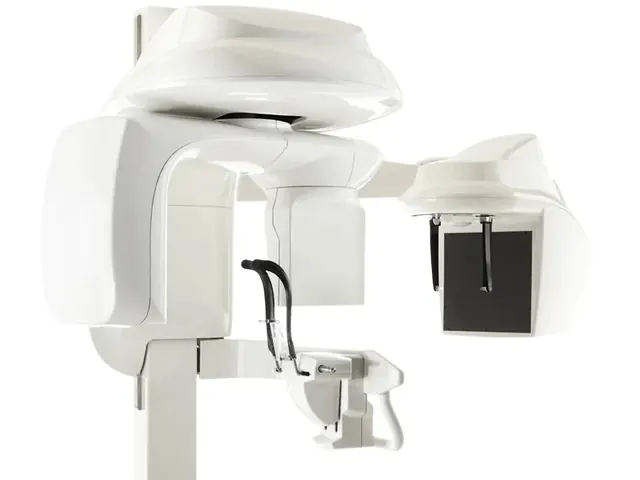
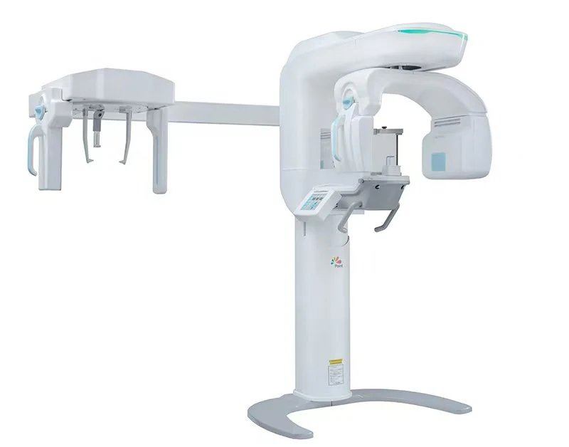
CBCT for temporomandibular joint (TMJ) imaging and upper airway / obstructive sleep apnea (OSA) assessment represents a specialty application niche at the intersection of dentistry, oral medicine, orthodontics, oral surgery, and sleep medicine. The increasing dental role in airway health and sleep-disordered breathing — particularly in mandibular advancement device therapy and myofunctional orthodontics — has driven demand for CBCT systems optimized for this application. This guide walks through CBCT selection for TMJ and airway imaging, clinical applications, and procurement considerations from Shanghai.
TMJ imaging clinical applications
TMJ morphology assessment
- Condyle morphology: condyle shape, size, cortical thickness bilateral assessment
- Articular eminence morphology: steepness and shape affects disc mechanics
- Glenoid fossa: depth and configuration
- Condyle-fossa relationship: centric position, joint space measurement
- Developmental asymmetry: bilateral comparison for asymmetry identification
Degenerative joint disease
- Osteoarthritis: condyle flattening, osteophytes, subchondral sclerosis, erosion
- Erosion patterns: generalized vs. focal, surface vs. subchondral
- Joint space narrowing: indicator of disc displacement or degeneration
- Staging degenerative changes: for treatment planning and progression monitoring
Trauma assessment
- Condyle fracture: fracture line identification, displacement assessment
- Fracture classification: intracapsular, condyle neck, subcondylar
- Surgical vs conservative decision support: based on fracture displacement and joint mechanics
- Healing assessment: follow-up imaging for bony union
Idiopathic condylar resorption (ICR)
- Progressive condylar volume loss
- Associated with ortho-surgical treatment in some patients
- Longitudinal monitoring
- Treatment decision support
Ankylosis assessment
- Bony ankylosis characterization
- Fibrous vs. bony ankylosis differentiation (combined with MRI)
- Surgical planning for arthroplasty
Pre-orthodontic TMJ assessment
- Screening before comprehensive orthodontic treatment
- Identification of pre-existing TMJ pathology
- Medico-legal baseline documentation
Pre-prosthetic occlusal rehabilitation
- Complex occlusal rehabilitation patients
- Assessment of joint capacity to accept occlusal changes
- Treatment planning for comprehensive occlusal reconstruction
CBCT protocol for TMJ
FOV
- Bilateral TMJ: 12×9cm or 12×6cm FOV capturing both joints
- Unilateral TMJ (targeted): 8×8cm FOV
- Large FOV for maxillofacial context: 16×10cm when combined with facial imaging
Voxel size
- 150µm: standard for TMJ imaging
- 100µm: high-resolution for fine erosive changes, trabecular detail
- 200µm: adequate for morphological assessment with lower dose
Positioning protocols
- Closed mouth: standard centric occlusion position
- Open mouth: for condyle translation assessment (separate scan)
- Bilateral simultaneous: both joints in single scan for comparison
Reconstruction considerations
- Bone window reconstruction: optimized for bony detail visualization
- Multi-planar reconstruction: axial, sagittal, coronal TMJ views
- Oblique sagittal reconstruction: aligned with individual condyle long axis for accurate assessment
- 3D volume rendering: for anatomic visualization and patient communication
Upper airway imaging clinical applications
Sleep apnea screening and evaluation
- Anatomical contributors to OSA: retropalatal narrowing, retrolingual narrowing, lateral pharyngeal wall
- Minimum cross-sectional area: predictor of OSA severity
- Airway volume: total upper airway volume measurement
- Baseline documentation: pre-treatment airway for monitoring post-treatment changes
- Note: CBCT alone does not diagnose OSA; diagnosis requires polysomnography. CBCT provides anatomical context.
Mandibular advancement device (MAD) treatment planning
- Pre-treatment airway mapping
- Treatment outcome verification (post-MAD advancement airway measurement)
- MAD optimization based on airway response
- Patient education and treatment acceptance
Airway-focused orthodontic treatment
- Myofunctional orthodontic approach integration
- Maxillary expansion airway outcomes
- Functional appliance airway outcomes
- Growth and development impact on airway
Pre-orthognathic surgical assessment
- Surgical planning considering airway impact
- Pre- and post-surgical airway comparison
- Setback vs. advancement surgical decisions with airway context
Pediatric airway assessment
- Adenotonsillar contribution to pediatric sleep-disordered breathing
- Growth-and-development airway monitoring
- Early intervention planning
CBCT protocol for airway imaging
FOV
- Large FOV: 16×10cm or 17×13cm capturing full upper airway from nasopharynx to hypopharynx
- Extended mandibular inclusion: airway from nasal choanae to epiglottis
Voxel size
- 200µm or 300µm: adequate for airway segmentation; dose-conscious for screening applications
- 150µm: when bone detail also needed (combined maxillofacial + airway)
- Dose priority: airway imaging typically screening; minimize dose while maintaining diagnostic capability
Patient positioning protocol
- Natural head position: seated or standing, looking straight ahead, relaxed
- No bite block: bite blocks can alter tongue position and mandibular position affecting airway
- End-expiration: standardized breathing position for reproducibility
- No swallowing during scan: operator instruction to patient before exposure
Airway segmentation software
- Automatic segmentation: algorithm identifies airway boundaries in CBCT volume
- Semi-automatic with manual correction: standard workflow for clinical use
- Volume and cross-sectional measurement: quantitative analysis of airway dimensions
- Color-coded 3D visualization: airway narrowing visualization for patient communication
- Software platforms: Dolphin Imaging airway module, Invivo, 3DSlicer (research), various specialized platforms
Combined TMJ + airway workflow
Many patients benefit from combined TMJ + airway assessment in single CBCT scan:
- Large FOV single scan: 16×10cm or 17×13cm captures both TMJs and full upper airway
- Efficient dose utilization: one scan serves both assessments vs. two separate exposures
- Clinical correlation: TMJ and airway are frequently both relevant (e.g. MAD therapy patients, TMD patients with breathing-related symptoms)
- Comprehensive maxillofacial context: whole craniofacial anatomy in single dataset
CBCT specifications for TMJ/airway specialty
- Large FOV capability: 16×10cm minimum, 17×13cm or larger preferred
- Dose-optimized large-FOV protocols: pulsed exposure or reduced mAs for airway screening
- Motion artifact robustness: longer scans more susceptible to patient movement; motion-robust reconstruction important
- Airway segmentation software: automatic or semi-automatic airway segmentation
- TMJ-specific protocols: bilateral symmetric capture with optimized positioning
- 3D rendering and analysis: quantitative measurement and visualization tools
Clinical workflow integration
Sleep medicine / MAD therapy workflow
- Initial sleep medicine evaluation (polysomnography)
- Referral to dental provider for MAD candidacy assessment
- CBCT for baseline airway mapping and anatomical suitability
- MAD fabrication and calibration
- Post-treatment polysomnography to confirm efficacy
- Follow-up CBCT (optional) to document airway response
TMD/TMJ specialty workflow
- Comprehensive TMJ examination (history, clinical exam)
- Imaging decision: CBCT for bony pathology, MRI for disc pathology (complementary)
- CBCT for TMJ morphology, degenerative changes, trauma
- Treatment planning: conservative, occlusal appliance, physical therapy, surgical
- Follow-up imaging for progression monitoring
Comprehensive airway-focused orthodontic workflow
- Initial orthodontic and airway evaluation
- CBCT for comprehensive craniofacial + airway assessment
- Treatment plan incorporating airway goals
- Progress CBCT (as indicated) for airway response monitoring
- Post-treatment documentation
Clinical economics
- CBCT scan fee (TMJ or airway): USD 150–450 per scan
- Specialty practice case volume: sleep dentistry practice 20–60 airway CBCT per month; TMJ specialty practice 15–40 TMJ CBCT per month
- MAD therapy revenue: USD 1,500–4,500 per device fabrication and titration
- Comprehensive treatment value: MAD therapy case with documented airway improvement substantially valued in sleep medicine context
- Payback: typical 18–36 months for dedicated sleep dentistry or TMJ specialty practice
Chinese CBCT options for TMJ/airway practice
Entry-tier large-FOV CBCT (USD 32,000–42,000 FOB)
- 16×10cm FOV capability
- 200µm voxel for airway, 150µm for TMJ
- Basic imaging software, DICOM export
- Fit: specialty practice commissioning CBCT for TMJ or airway focus
Mid-tier TMJ/airway CBCT (USD 42,000–58,000 FOB)
- 17×13cm FOV capability
- Dose-optimized protocols for airway screening
- Integrated airway segmentation software
- TMJ-specific reconstruction protocols
- Fit: mainstream specialty practice
Premium specialty CBCT (USD 58,000–80,000 FOB)
- Premium large-FOV capability with optimized image quality
- Advanced airway and TMJ analysis software
- Integration with sleep medicine workflow tools
- Fit: specialty sleep dentistry / TMJ center, academic institution
Limitations of CBCT for TMJ/airway
TMJ
- CBCT shows bone but not disc or soft tissue; MRI required for complete TMJ assessment
- Dynamic TMJ motion not captured in static CBCT; functional imaging alternatives needed for some questions
- Early degenerative changes may be more sensitively detected on MRI
Airway
- Static CBCT captures single moment; dynamic airway changes during sleep not captured
- CBCT not a substitute for polysomnography in OSA diagnosis
- Head position standardization critical for reproducibility
- Supine vs. seated airway differs; CBCT upright position differs from sleep position
Regulatory considerations
- Classification: Class IIb medical device (CE-MDR); Class II 510(k) FDA
- Radiation safety: destination country radiation safety framework
- Airway analysis software: Class IIa medical device software if quantitative output used for clinical decision-making
- Sleep medicine integration: collaboration with sleep medicine specialists; understand local regulatory framework for dental role in sleep medicine
Sourcing CBCT for TMJ or airway specialty practice?
WhatsApp us with your specialty focus (TMJ/TMD, sleep dentistry/MAD, airway-focused orthodontic, comprehensive maxillofacial), expected monthly specialty CBCT volume, interest in airway segmentation software and TMJ-specific analysis tools, destination country, and budget. We’ll propose CBCT options with large FOV capability and dose-optimized specialty protocols matched to your workflow, discuss combined TMJ + airway workflow strategy, and quote FOB Shanghai pricing with full commissioning package landed cost analysis.
Ready to price this out?
Tell us the model, quantity and destination port. We reply with FOB or CIF pricing, current stock status and a video demo where the unit is in our Shanghai warehouse.